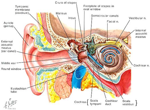
1. Sound waves enter the outer ear and travel through the external auditory meatus or ear canal, which leads to the eardrum.
2. The eardrum or tympanic membrane vibrates from the incoming sound waves and sends these vibrations to three tiny bones in the middle ear. These bones are called the malleus, incus, and stapes.
3. The bones in the middle ear amplify, or increase, the sound vibrations and send them to the cochlea, a snail-shaped structure filled with fluids in the inner ear. An elastic partition runs from the beginning to the end of the cochlea, splitting it into an upper and lower part. This partition is called the basilar membrane upon which key structures sit.
4. Once the vibrations cause the fluid inside the cochlea to ripple, a traveling wave forms along the basilar membrane. Sensory hair cells sitting on top of the basilar membrane – ride the wave. Sensory hair cells near the wide end of the cochlea detect higher pitched sounds, such as an infant crying. Those close to the center detect lower pitched sounds, such as a large dog barking.
5. As the sensoriy hair cells move, microscopic hair-like projections (known as stereocillia) that perch on top of the hair cells bump against an underlying structure and bend. Bending causes pore-like channels, shich are at the tips of the stereocilia, to open up. When that happens, chemicals rush into the cells, creating an electrical signal.
6. The auditory nerve carries the signal through the brain stem and up to the auditory cortex, which turns into sounds and speech that we understand.